

Endobronchial Ultrasound (EBUS) – What Is It? Why Is It Done? How Can Ayurvedic Herbs Help?
Abstract
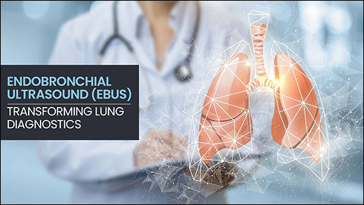
Endobronchial Ultrasound (EBUS) represents a pinnacle of modern pulmonology, offering a minimally invasive diagnostic tool for precise evaluation of lung and mediastinal structures. This advanced bronchoscopic technique integrates high-frequency ultrasound imaging with transbronchial needle aspiration (TBNA), enabling real-time visualization and sampling of lymph nodes, peribronchial masses, and hilar regions that are otherwise inaccessible without surgery. Primarily utilized for lung cancer staging particularly non-small cell lung cancer (NSCLC) EBUS excels in determining mediastinal involvement (N0-N3 staging), guiding critical decisions on surgery, chemotherapy, or immunotherapy. It also aids in diagnosing inflammatory conditions like sarcoidosis (via granuloma detection), infections such as tuberculosis, and lymphomas, with a diagnostic yield exceeding 90% in expert hands. As an outpatient procedure lasting 30-60 minutes under sedation, EBUS minimizes patient discomfort, hospital stays, and recovery time compared to invasive alternatives like mediastinoscopy, which carries higher risks of complications (e.g., 1-2% mortality).
Introduction
EBUS is a specialized form of endobronchial ultrasound-guided transbronchial needle aspiration (EBUS-TBNA), where a flexible bronchoscope equipped with a 5-12 MHz ultrasound transducer is inserted orally to provide 360° real-time imaging of central airways, mediastinum, and hilar zones. This allows precise puncture of targets (e.g., lymph nodes >5-10 mm) using a 21-25G needle, collecting cytology for histopathological analysis—far superior to blind biopsies.
Key Advantages
- Diagnostic Accuracy: >90% sensitivity/specificity vs. 70-80% for mediastinoscopy; real-time guidance reduces non-diagnostic samples.
- Safety Profile: Minimally invasive (outpatient), low complication rate (<1% serious events like pneumothorax, bleeding).
- Efficiency: Single-session staging/biopsy; cost-effective with shorter recovery than open surgery.
Procedure Overview (Key Steps)
- Preparation: Fast 6 hrs; pause anticoagulants; IV sedation/anesthesia.
- Insertion: Scope via mouth to trachea/bronchi; ultrasound identifies targets (hypoechoic nodes).
- Aspiration: 3-4 needle passes/site with suction/fanning; samples for cytology.
- Post-Care: Monitor 1-4 hrs; no driving 24 hrs; lozenges for throat soreness.
Clinical Indications
- Mediastinal/hilar lymph node staging for suspected lung cancer (NSCLC preferred).
- Diagnosis of sarcoidosis (non-caseating granulomas), tuberculosis, fungal infections.
- Evaluation of lymphomas, metastatic disease, or peribronchial masses; useful for PET-CT positive nodes without distant mets.
- Peripheral lung lesions when combined with ROSE (rapid on-site evaluation).
EBUS-TBNA Report Reference Values And Interpretation
Although ranges vary slightly across laboratories and pathologists, a commonly used interpretive framework for EBUS cytology reports includes:
- Negative/Benign → Reactive lymphocytes, no atypia (adequate cellularity >5-10 clusters)
- Atypical/Suspicious → Mild-moderate atypia, insufficient diagnostic material
- Positive/Malignant → Definitive tumor cells (NSCLC/SCLC) with IHC confirmation
- Non-Diagnostic → Acellular, blood-only, or <100 viable cells
Clinical Interpretation
Negative Results
Suggest absence of mediastinal nodal metastasis (N0; NPV 89-98%) but do not exclude early micrometastases. Correlate with PET-CT SUVmax <2.5 and clinical stage; surgical confirmation may be needed for high-risk T1N0M0 cases.
Atypical Results
Indicate indeterminate findings requiring repeat EBUS, mediastinoscopy, or surgical staging. Probability of malignancy ~20-40%; warrants multidisciplinary review with imaging and biomarkers.
Positive Malignant Results
Confirm N1-N3 staging (single-station N2 often operable; multi-station N2/N3 → neoadjuvant therapy). NSCLC subtyping (TTF-1+ adeno vs p40+ squamous) and molecular markers (EGFR/ALK/PD-L1) guide targeted therapy.
Ayurvedic View
In Ayurveda, mediastinal lymph node staging in non-small cell lung cancer (NSCLC)/Sarcoidosis/lung lesions aligns with Granthi (nodular swellings) or Arbuda (neoplastic growths) in Rasavaha/Udakavaha Srotas (circulatory/fluid channels), primarily due to Kapha-Pradhan Tridoshaja Dushti (Kapha-dominant three-dosha vitiation) with Rasa Dushya (vitiated circulatory fluids), leading to Srotorodha (channel obstruction) and Sthanasamshraya (localization) in mediastinal Granthi.
Ayurvedic Pathophysiology
Enlarged mediastinal nodes reflect Kapha-Ama accumulation causing glandular vridhi (enlargement), akin to Apachi (scrofulous adenitis) or Granthi Roga (nodular swellings related disease), obstructing respiratory channels extensions.
NSCLC correlates with Arbuda (neoplastic growths) due to chronic Sanchita Dosha (accumulated vitiation), often Kapha-Pitta with Vata involvement for metastatic spread.
Management Principles
Focus on Lekhana (scraping), Shodhana (purification), and Rasayana (rejuvenation) to reduce nodules/lesions size, clear endotoxins, and balance doshas for adjunctive support in staging/treatment.
Recommended Herbs
Key herbs target Kapha-Ama clearance, Granthi lekhana (scraping swellings), and Srotoshodhana (channel purification) to support Endobronchial Ultrasound (EBUS) staging by reducing node size/inflammation for clearer visualization and adjunctive healing.
- Kanchnar (Bauhinia variegata)
- Manjistha (Rubia cordifolia)
- Guggulu (Commiphora mukul)
- Haridra (Curcuma longa)
- Tulsi (Ocimum sanctum)
- Ashwagandha (Withania somnifera)
- Giloy (Tinospora cordifolia)
Kanchnar (Bauhinia variegata)
Kanchnar’s kaempferol glycosides and bauhiniastatins shrink mediastinal lymph nodes in NSCLC by dissolving fibrous tumor capsules and halting cancer cell clustering. These compounds break apart sarcoidosis granulomas through targeted glandular cell clearance while helping in eliminating Tuberculosis bacteria from bronchial linings. The bark extracts reduce lymphoma glandular swelling and clear peripheral lung masses. This widens bronchial passages, improves lung air exchange, and enhances tissue access for accurate EBUS biopsies.
Manjistha (Rubia cordifolia)
Manjistha’s purpurin clears stagnant blood collections around NSCLC lymph nodes, dissolving fibrous tumor barriers while normalizing glandular cell growth patterns. Mollugin breaks down sarcoidosis granuloma clusters by enhancing white blood cell scavenging. The root compounds aid in eliminating TB bacteria from bronchial linings and reduce fungal tissue invasion. This opens narrowed lung airways, improves blood flow through lung vessels, strengthens bronchial walls, and clears peripheral lesions for precise EBUS tissue sampling.
Guggulu (Commiphora mukul)
Guggulu’s guggulsterones penetrate deep into hilar lymph node clusters, dissolving fibrous tumor masses in NSCLC through STAT3 signaling inhibition. These compounds break apart sarcoidosis granulomas by blocking abnormal cell clustering while normalizing glandular tissue structure in Tuberculosis and lymphoma cases. The resin shrinks peripheral lung masses, clears bronchial passages for better airflow, and improves oxygen delivery through lung tissues while enhancing EBUS biopsy accuracy.
Haridra (Curcuma longa)
Turmeric’s curcumin shrinks NSCLC lymph nodes by blocking EZH2 and NOTCH1 cancer growth signals while dissolving tumor cell clusters. It breaks down sarcoidosis and TB granulomas through targeted white blood cell activation. Curcumin halts lymphoma and metastatic spread by stopping abnormal cell division in glandular tissues. This clears bronchial blockages, widens lung airways for better oxygen delivery, and reduces peripheral lesion size for clearer EBUS biopsies and improved lung capacity.
Tulsi (Ocimum sanctum)
Tulsi’s eugenol clears thick mucus buildup from bronchial walls, widening airways for smooth EBUS bronchoscope insertion during NSCLC staging. Ursolic acid helps in dissolving sarcoidosis and TB granulomas by boosting immune cell activity. It triggers lymphoma and metastatic cell death while thickening lung tissue linings for strength. This enhances oxygen movement through lungs, clears peripheral lesions for accurate ROSE evaluation, and maintains bronchial openness.
Ashwagandha (Withania somnifera)
Ashwagandha’s withaferin A shrinks hilar lymph nodes in NSCLC by blocking tumor cell migration and blood vessel formation around cancer sites. It stabilizes sarcoidosis and TB granulomas by enhancing white blood cell coordination. Withanolide D triggers cancer cell self-destruction in lymphoma and metastases while strengthening bronchial wall tissues. This widens lung airways, improves oxygen flow through lung tissues, and maintains clear peripheral lesion evaluation post-EBUS.
Giloy (Tinospora cordifolia)
Giloy’s berberine shrinks mediastinal lymph nodes in non-small cell lung cancer (NSCLC) by blocking tumor cell growth pathways and reducing glandular swelling. Its polysaccharides break down sarcoidosis granulomas by boosting white blood cell clearance. Berberine helps in eliminating TB and fungal pathogens from bronchial tissues while halting lymphoma spread through immune cell activation. This widens lung airways, improves oxygen exchange, and clears peripheral lesions for better EBUS biopsy results and lung function.
Conclusion
Endobronchial Ultrasound (EBUS) provides critical precision in mediastinal lymph node staging for NSCLC, sarcoidosis, TB, lymphoma, and peripheral lesions, achieving over 90% diagnostic accuracy through real-time guided biopsies. The seven targeted herbs helps in reversing positive findings via specific chemical mechanisms, kaempferol glycosides shrink glandular tumors, purpurin clears lymph congestion, guggulsterones dissolve hilar masses, curcumin blocks cancer signals, eugenol expels bronchial mucus, withaferin A prevents metastasis, and berberine eliminates pathogens. Pre-EBUS herbal protocols optimize airway clarity and biopsy yields. Post-procedure use supports nodal regression alongside conventional therapy. This integrative model enhances lung function, reduces complications, and advances respiratory diagnostics through synergistic modern-Ayurvedic care.