

CT Enterography – What Is It? Why Is It Done? How Can Ayurvedic Herbs Help?
Abstract
In the landscape of the Mahasrotas, Crohn’s disease manifests as a turbulent storm of Vata and Pitta, where the sacred fire of Agni turns destructive. This chronic inflammatory odyssey transcends mere tissue damage, representing a profound Dhatu Paka that erodes the Mamsadhara Kala. From the "Comb Sign" of engorged vessels to the scarring of fibrotic strictures, the body’s internal channels (Srotas) lose their fluidity, replaced by the rigidity of Sanga. It is a clinical dance of deep-seated Ama, where the intelligence of the gut is compromised, requiring a harmonious integration of modern diagnostics and ancient restorative wisdom. CT Enterography emerges as the diagnostic gold standard, utilizing high-resolution neutral contrast to illuminate the hidden architecture of the small bowel. It precisely identifies active inflammation, fibrotic strictures, and mesenteric vascular engorgement, providing a definitive map of Srotodushti for clinical intervention.
Introduction
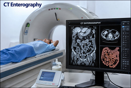
CT Enterography (CTE) is a specialized computed tomography scan designed to produce high-resolution, cross-sectional images of the small intestine. Unlike a standard abdominal CT, CTE uses a large volume of neutral oral contrast (such as polyethylene glycol or mannitol) to distend the bowel loops, followed by intravenous (IV) contrast to highlight the intestinal wall and blood vessels. This technique is the gold standard for diagnosing and monitoring Crohn’s disease, as it accurately detects wall thickening, ulcers, and complications like fistulas or abscesses. It is also highly effective for identifying small bowel tumors, obstructions, and sources of internal bleeding.
What Happens During The Procedure?
During a CT Enterography (CTE), the procedure is divided into a preparation phase and the actual scanning phase. The goal is to fully distend the small intestine to allow for a clear view of the bowel wall.
Phase 1: Oral Preparation
- Arrival: You will typically arrive 60 to 90 minutes before the scan.
- Contrast Consumption: You will be asked to drink a large volume (usually 1 to 1.5 liters) of a neutral oral contrast solution.
- Timing: This is consumed at a steady pace over the hour leading up to the scan. This solution does not get absorbed quickly, ensuring the entire length of the small bowel is "inflated" and visible.
Phase 2: The Scan
- Positioning: You will lie flat on the CT table. A technician may place an IV line in your arm.
- IV Contrast: Just before the scan begins, an iodinated IV contrast is injected. You might feel a brief warm sensation or a metallic taste in your mouth; this is normal.
- Scanning: The table moves through the doughnut-shaped scanner. You will be asked to hold your breath for a few seconds at a time to ensure the images are crisp and not blurred by movement.
- Duration: The actual imaging takes less than 10 to 30 seconds, though the total time in the room is about 10 to 15 minutes.
Phase 3: Post-Procedure
- Hydration: You will be encouraged to drink plenty of water to help flush the IV contrast through your system.
- Side Effects: Because of the large volume of oral contrast, it is common to experience a mild laxative effect shortly after the procedure.
Indications
CT Enterography (CTE) is the preferred imaging modality for evaluating the small bowel, particularly when there is a need to visualize the bowel wall and the surrounding blood vessels simultaneously. The primary clinical indications include:
1. Inflammatory Bowel Disease (IBD)
- Crohn’s Disease: This is the most common indication. CTE is used to diagnose the disease, assess its activity (flare-ups), and identify complications such as fistulas, abscesses, or phlegmon.
- Strictures: Identifying narrowed areas of the bowel that may cause chronic pain or obstruction.
2. Small Bowel Neoplasms
- Tumors: Detecting and staging benign or malignant growths, such as adenocarcinomas, carcinoid tumors, or lymphomas, which can be difficult to see on standard CT scans.
- Polyps: Screening in patients with hereditary syndromes like Peutz-Jeghers.
3. Gastrointestinal Bleeding
- Obscure GI Bleeding: When a colonoscopy and upper endoscopy are negative, CTE can locate bleeding sources in the small intestine, such as angiodysplasia or small ulcers.
4. Bowel Obstruction and Ischemia
- Partial Obstruction: Evaluating the cause and location of a blockage.
- Mesenteric Ischemia: Assessing blood flow to the intestines in cases of suspected "abdominal angina" or vascular insufficiency.
5. Malabsorption and Celiac Disease
- Evaluating structural changes or complications in patients with non-responsive Celiac disease or unexplained weight loss.
Results Interpretation
Interpreting the results of a CT Enterography involves a systematic evaluation of the bowel wall, the lumen (the space inside the bowel), and the surrounding structures like blood vessels and lymph nodes.
|
Finding |
Description |
Clinical Significance |
|
Mural Hyper-enhancement |
The bowel wall appears abnormally bright after IV contrast. |
Indicates active inflammation (e.g., acute Crohn’s flare). |
|
Wall Thickening |
Bowel wall measures > 3mm in a distended segment. |
Common in IBD, ischemia, or intramural hemorrhage. |
|
Mural Stratification |
A "target" or "halo" appearance of the intestinal layers. |
Suggests mural edema; helps distinguish active from chronic disease. |
|
Comb Sign |
Engorged, prominent vasa recta (blood vessels) near the bowel. |
A specific sign of active, advanced inflammation in the mesentery. |
|
Luminal Narrowing |
Reduced diameter of the bowel lumen. |
May indicate a stricture (fibrotic/permanent or inflammatory/reversible). |
|
Pre-stenotic Dilation |
Widening of the bowel segment before a narrowing. |
Indicates a hemodynamically significant obstruction. |
|
Sacculations |
Outpouchings on the border of the bowel. |
Often seen in chronic Crohn’s disease due to asymmetrical scarring. |
Ayurveda’s Insight
In the context of CT Enterography, Ayurveda provides a fascinating lens through which we can interpret the structural and functional changes seen on the scan. While a CT scan identifies Rupa (the manifested form), Ayurvedic principles help categorize the underlying Samprapti (pathogenesis).
Pros And Cons Of Optical Coherence Tomography
1. Structural Changes as Dhatu Paka and Srotodushti
The high-resolution images of the bowel wall capture the progression of Dhatu Paka (tissue inflammation/metabolic breakdown).
- Mural Hyper-enhancement: Correlates with Pitta Vidagdha (acute inflammatory heat) within the Mamsa (muscle) and Rakta (blood) layers of the gut.
- Wall Thickening/Edema: This is a clear manifestation of Tarpaka Kapha or Kleda (excess moisture/congestion) causing obstruction within the Annavaha Srotas (digestive channels).
2. The “Comb Sign” and Siragranthi
The Comb Sign (engorged vasa recta) seen on CTE is a physical representation of Vata-Rakta or Raktagata Vata.
- In Ayurveda, when Vata is aggravated, it pushes the Rakta Dhatu into the smaller Siras (vessels), causing the engorgement and tortuosity seen on the scan. This is often an indication of Udaana or Samana Vata dysfunction.
3. Strictures and Sanga (Obstruction)
A stricture or pre-stenotic dilation is a classic example of Sanga (blockage) in the Srotas.
- Fibrotic Strictures: These represent a state of Dhatu Kshaya (tissue depletion) and chronic Vata dryness, where the supple Mamsadhara Kala (mucous membrane) has become hard and scarred (Khara guna).
- Inflammatory Strictures: These are more indicative of Ama (toxic accumulation) and Kapha-Pitta blockage, which may still be reversible with Deepana-Pachana (digestive stimulant) therapies.
Now We Will Talk About Some Herbs That Support Management Of Crohn’s Disease
1. Kutaj (Holarrhena antidysenterica)
Kutaj is pharmacologically prized for its potent alkaloids, primarily conessine. It exhibits significant antiprotozoal and antibacterial activity, specifically targeting Entamoeba histolytica. It acts as a natural astringent on the intestinal mucosa, reducing excessive fluid secretion and hypermotility. Known as the premier Arshoghna (anti-hemorrhoid) and Kutaj-ghan (anti-diarrheal), it possesses Tikta (bitter) and Kashaya (astringent) Rasa with a Sheetal (cooling) Veerya. It absorbs excess Kleda (moisture) and balances Pitta-Kapha in the Annavaha Srotas. In Crohn’s, Kutaj helps arrest chronic diarrhea and controls the bleeding associated with Raktatisara (ulcerative/bloody stools). By strengthening the mucosal barrier, it prevents the "leakage" of fluids and toxins into the intestinal lumen.
2. Bilva (Aegle marmelos)
Bilva contains marmelosin and tannins. Its unique pharmaceutical property lies in its dual action: the unripe fruit is an astringent for diarrhea, while the ripe fruit acts as a laxative. It exhibits strong anti-inflammatory properties, protecting the gut from oxidative stress and regulating the gut-brain axis. Highly valued for its Grahi (absorbent) and Deepana (digestive-kindling) properties. It is Ushna (heating) in Veerya and has a Kashaya-Tikta Rasa. It is specifically indicated for Grahani (malabsorption) by improving the "holding" capacity of the intestines. Bilva is essential for managing the Grahani aspect of Crohn’s. It helps the intestines properly process nutrients and prevents the passage of undigested food (Ama), which is a primary trigger for inflammation and bowel urgency.
3. Arjuna (Terminalia arjuna)
Arjuna is rich in arjunolic acid and flavonoids. While famous as a cardiotonic, it is scientifically recognized as a potent hemostatic (stops bleeding) and wound-healing agent. It promotes collagen synthesis and strengthens the endothelial lining of blood vessels, reducing vascular permeability. Described as Sandhaniya (tissue-joining) and Hridya. It has a dominant Kashaya (astringent) Rasa and Sheetal (cool) Veerya. It is a powerful Rakta-Stambhana that helps heal internal Kshata (wounds/ulcers) within the Srotas. For Crohn’s patients, Arjuna assists in the repair of deep intestinal ulcers. Its astringent nature helps close the "micro-wounds" in the bowel wall, reducing the risk of protein loss and internal hemorrhaging often seen on CT Enterography as wall friability.
4. Pitapapda (Fumaria indica)
Pitapapda contains alkaloids like protopine. It is scientifically known for its hepatoprotective and cholagogue actions, stimulating bile flow and detoxifying the liver. It has antipyretic properties and acts as a systemic blood purifier, reducing pro-inflammatory cytokines in the bloodstream. Known as Parpata, it is the ultimate Jwaraghna (fever-remover). It is Tikta (bitter) and Sheeta (cold). It acts as a Rakta-Prasadana (blood cleanser), removing Pitta toxins from the Rakta Dhatu and cooling the body’s internal temperature. During a Crohn’s flare, systemic inflammation often leads to low-grade fever and metabolic heat. Pitapapda cools the "heat" in the blood, reducing the systemic inflammatory response and clearing the Ama that fuels the autoimmune-like attack on the gut.
5. Patol (Trichosanthes dioica)
Pointed Gourd is a nutrient-dense botanical containing linoleic acid and antioxidants. It acts as a potent gastric sedative and hepatoprotective agent. It improves peristalsis without causing irritation and stimulates the gall bladder to release bile, ensuring smoother fat digestion and metabolic balance. Regarded as the most Pathya (wholesome) vegetable for the gut. It is Tikta (bitter) yet Hridya (pleasing) and Deepana. It is Tridosha-shamaka, meaning it balances all three Doshas, particularly clearing Srotas blockages and Patol is the "safe" fiber for Crohn’s. It clears the Srotodushti (channel blockage) and Sanga seen in strictures without aggravating the bowel wall. It provides a gentle way to kindle Agni while soothing the inflamed Pitta environment of the intestines.
Conclusion
At last it can be concluded that the integration of CT Enterography with Ayurvedic therapeutics represents a paradigm shift in managing Crohn’s disease, moving from symptomatic relief to deep structural healing. While CTE provides the "visual evidence" of Srotodushti—mapping the precise location of inflammation and strictures—Ayurveda offers the "functional cure" to restore the Mamsadhara Kala. By utilizing botanicals like Kutaj, Bilva, and Arjuna, we can systematically neutralize Ama, cool Pitta-led inflammation, and repair the intestinal lining. This synergy ensures that modern diagnostics guide the application of ancient wisdom, ultimately achieving the shared goal of long-term mucosal integrity and systemic balance.