

Optical Coherence Tomography (OCT) – What Is It? Why Is It Done? How Can Ayurvedic Herbs Help?
Abstract
The human eye is a silent storyteller, a shimmering archive of our most delicate health secrets. Optical Coherence Tomography acts as the ultimate visionary translator, peeling back the invisible curtains of the retina to reveal a microscopic landscape of light and shadow. Like an "optical biopsy," this test captures the silent architecture of sight—mapping the golden peaks of the macula and the deep valleys of the optic nerve without ever touching the surface. It is where ancient observation meets futuristic precision, turning pulses of near-infrared light into a roadmap for preservation, ensuring the windows to our soul remain clear, vibrant, and eternally bright.
Introduction
Optical Coherence Tomography (OCT) is a non-invasive, high-resolution imaging technique that functions similarly to "ultrasound with light." Instead of sound waves, it uses low-coherence light (typically near-infrared) to capture micrometer-resolution, cross-sectional images of biological tissue. By measuring the echo delay and intensity of backscattered light, it creates detailed 3D reconstructions of internal structures without requiring physical contact or ionizing radiation.The significance of OCT lies in its ability to provide "optical biopsies," allowing clinicians to see beneath the surface of tissues in real-time. It has become the gold standard in ophthalmology for diagnosing and managing retinal diseases like glaucoma and macular degeneration. Beyond eye care, its prevalence is rapidly expanding into cardiology (to visualize arterial plaque) and dermatology, making it one of the most vital diagnostic tools in modern precision medicine.
What Happens During The Procedure?
The procedure for an Optical Coherence Tomography (OCT) scan is remarkably straightforward, quick, and painless. It usually takes about 5 to 10 minutes in total.
1. Preparation
In some cases, your doctor may use dilating eye drops to widen your pupils, making it easier to examine the retina. However, many modern OCT machines can capture high-quality images without dilation.
2. Positioning
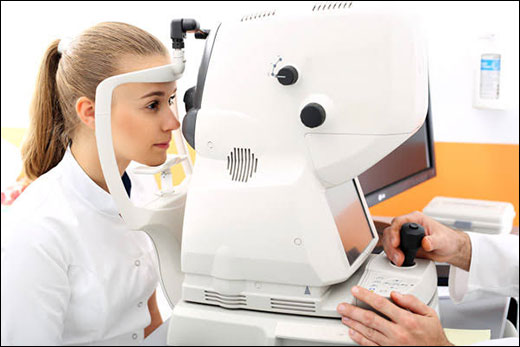
You will sit in front of the OCT machine and rest your chin on a padded support and your forehead against a bar to keep your head perfectly still.
3. The Scan
You will be asked to stare at a target light inside the machine (usually a small green cross or dot). The equipment will then scan your eye using a beam of light. You won’t feel anything, and there is no physical contact with your eye.
4. Data Capture
The machine uses interferometry to measure the light reflections. It creates a "map" of the different layers of the retina or the optic nerve in real-time.
Indications of Optical Coherence Tomography
OCT is a versatile tool used across several medical fields, though it is most renowned in ophthalmology. Its primary indications are for conditions where high-resolution, cross-sectional imaging of thin tissue layers is required.
1. Ophthalmology (Primary Use)
This is the "gold standard" for diagnosing and monitoring diseases of the retina and optic nerve, including:
- Macular Degeneration (AMD): To detect fluid leakage or "drusen" (deposits) under the retina.
- Glaucoma: To measure the thickness of the Retinal Nerve Fiber Layer (RNFL); thinning often indicates early nerve damage.
- Diabetic Retinopathy: To identify swelling (Macular Edema) and leaking blood vessels.
- Macular Holes & Pucker: To visualize physical tears or tension (traction) on the macula.
- Retinal Detachment: To see where the retina has lifted away from its underlying support.
2. Cardiology
Intravascular OCT (IV-OCT) is used during cardiac catheterization to:
- Plaque Characterization: Identify "vulnerable plaques" (lipid-rich areas with thin caps) prone to causing heart attacks.
- Stent Optimization: Ensure coronary stents are properly expanded and opposed against the vessel wall.
3. Dermatology & Other Fields
- Skin Cancer: Assessing margins of basal cell or squamous cell carcinomas without an immediate surgical biopsy.
- Gastroenterology: Imaging the esophagus (e.g., Barrett’s esophagus) to detect early precancerous changes.
Pros And Cons Of Optical Coherence Tomography
|
Pros (Advantages) |
Cons (Limitations) |
|
Non-Invasive: No needles, radiation, or physical contact. |
Low Penetration: Light only reaches 2–3 mm deep. |
|
Micro-Detail: Sees tissue layers at a microscopic level. |
Blocked by Opacity: Cannot see through cataracts or blood. |
|
Ultra-Fast: Complete 3D scans in just seconds. |
Stability Needed: Patient must sit perfectly still. |
|
Precise Tracking: Perfect for monitoring disease over time. |
High Cost: The equipment is very expensive. |
Results Interpretation
Physician interpret OCT results using two main views:
1. The Cross-Section (The “Image”)
Normal:
- Smooth, distinct layers with a central dip (the fovea).
Abnormal:
- Dark pockets: Indicate fluid or swelling (edema).
- White lumps: Indicate deposits or scarring (drusen).
- Lifting/Tears: Indicate the retina is pulling away.
2. The Color Map (The "Traffic Light")
The machine compares your eye to a database of healthy people:
- Green: Normal thickness (Healthy).
- Yellow: Borderline thin (Warning).
- Red: Abnormally thin (Potential damage/disease)
Ayurveda’s Insight!
Ayurveda views the eye as the seat of Alochaka Pitta (the fire of vision), and modern Optical Coherence Tomography scans help visualize its balance. When an Optical Coherence Tomography scan shows thinning or atrophy (Red zones), it correlates with a Vata imbalance or a loss of Majja Dhatu (nerve tissue). Conversely, fluid pockets or swelling (Edema) are seen as a Kapha blockage, while bleeding or active inflammation indicates aggravated Pitta. By using Optical Coherence Tomography to identify these "energetic" signatures, Ayurvedic practitioners can tailor traditional therapies like Netra Tarpana (medicated ghee pooling) to nourish Vata-related thinning or Seka (medicated pouring) to clear Kapha-related fluid, effectively bridging ancient wisdom with high-tech diagnostic precision.
1. Amla (Emblica officinalis)
Known as a potent Rasayana (rejuvenative), Amla is specifically "Chakshushya" (strengthening to the eyes). It balances all three Doshas, particularly Pitta, which governs vision. It is believed to cool the eyes and prevent "burning" sensations. Amla is one of the richest natural sources of Vitamin C and polyphenols. These antioxidants neutralize free radicals that cause oxidative stress in the retina. Studies suggest it helps prevent cataracts by inhibiting glucose-induced protein crystallization and protects against Optical Coherence Tomography-detected retinal thinning.
2. Gotukola (Centella asiatica)
Often called Mandukaparni, it is prized for its ability to nourish the Majja Dhatu (nervous system). It improves the “Prana” or life force flowing to the sensory organs, specifically helping with mental clarity and visual focus. Gotu Kola enhances microcirculation and strengthens the walls of small blood vessels (capillaries). This is vital for conditions like diabetic retinopathy, where vessel leakage is common. It contains triterpenoids that support collagen synthesis, maintaining the structural integrity of the eye’s connective tissues and the optic nerve.
3. Bhringraj (Eclipta alba)
Known as the "Ruler of the Hair," it is equally powerful for the eyes (Netya). It is used to detoxify the liver, which Ayurveda closely links to eye health. By balancing Ranjaka Pitta, it ensures the blood flowing to the eyes is pure and nutrient-rich. Research highlights its high content of wedelolactone and flavonoids, which provide potent anti-inflammatory and hepatoprotective effects. By reducing systemic inflammation, it helps manage chronic inflammatory eye conditions like uveitis and protects the retinal cells from premature aging and degeneration.
4. Bilberry (Vaccinium myrtillus)
While not native to ancient India, it is classified by modern Ayurvedic practitioners as a cooling, Pitta-pacifying fruit that improves the “Tejas” (radiance) of the eyes. It is often used as a modern supplement to traditional Rasayana therapies. Bilberry is famous for its high concentration of anthocyanosides. These pigments accelerate the regeneration of rhodopsin, the purple pigment used by rods in the eye for night vision. It also strengthens retinal capillaries, making it a primary defense against macular degeneration and glaucoma progression.
5. Triphala (The Three Fruits)
A cornerstone of eye care, this formula (Amalaki, Bibhitaki, Haritaki) is the ultimate bowel and tissue cleanser. It ensures that toxins (Ama) do not accumulate in the ocular channels. When used as a wash or taken internally, it balances the entire body to support the eyes. Triphala provides a synergistic blend of Vitamin C, tannins, and gallic acid. It has been scientifically shown to exhibit anti-glycation properties, which is crucial for preventing lens opacification (cataracts). It also helps regulate intraocular pressure, making it a valuable adjunct in managing Glaucoma.
Conclusion
Finally, It can be said that Optical Coherence Tomography stands as a revolutionary bridge between advanced physics and clinical care, transforming how we "see" the human eye. By utilizing low-coherence light to provide microscopic, cross-sectional "optical biopsies," it has turned the once-hidden layers of the retina into an open book. This non-invasive, high-speed diagnostic tool is indispensable for the early detection of silent sight-stealers like Glaucoma and Macular Degeneration, offering a level of precision that standard exams simply cannot match. When viewed through the timeless lens of Ayurveda, these technological insights gain a deeper dimension. The thinning or swelling captured on a scan becomes a roadmap for balancing the Doshas—Vata, Pitta, and Kapha—allowing for a truly holistic approach to vision. By combining the digital accuracy of Optical Coherence Tomography with the nourishing power of Chakshushya herbs like Triphala, Amla, and Bilberry, we can move beyond mere diagnosis toward a future of proactive, personalized eye preservation.