


Hyperpolarized Gas MRI – What Is It? Why Is It Done? How Can Ayurvedic Herbs Help?
Abstract
Assessment of regional lung ventilation and perfusion is central to the diagnosis, monitoring, and management of chronic respiratory disorders like COPD, asthma, and pulmonary fibrosis. Conventional imaging such as CT or V/Q scans provides structural or mismatched perfusion data but often involves radiation and lacks direct quantification of gas exchange or alveolar microstructure. Non-invasive functional tests that map ventilation defects, diffusion capacity, and blood-gas transfer offer critical clinical insight. Hyperpolarized Gas MRI was developed using polarized noble gases like Xenon-129 (¹²⁹Xe) or Helium-3 (³He) to provide high-resolution, radiation-free imaging of lung physiology. By evaluating gas distribution, apparent diffusion coefficients, and dissolved-phase transfer to tissue/red blood cells, the technique reflects real-time regional pulmonary performance.
Introduction
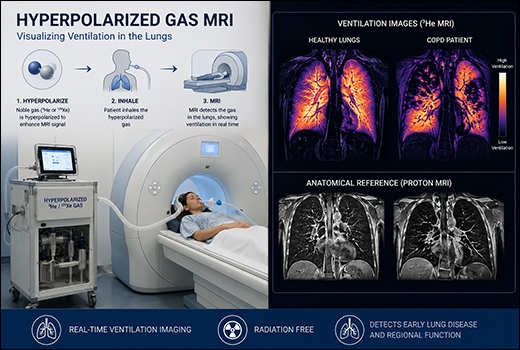
Hyperpolarized Gas MRI enhances lung imaging by using polarized noble gases, inhaled during breath-holds to produce high-contrast images of ventilation, perfusion, gas exchange, and microstructure. This investigational technique surpasses conventional MRI limitations in air-filled lungs, offering non-invasive, radiation-free assessment of regional function in diseases like COPD, Asthma, and Idiopathic Pulmonary Fibrosis.
Key Applications
It excels in static/dynamic ventilation mapping to quantify defects and heterogeneity, diffusion-weighted imaging for alveolar size (ADC values), and dissolved-phase ¹²⁹Xe for gas transfer to barriers/red blood cells, aiding precision in bronchodilator response or transplant planning. Perfusion can be evaluated via V/Q matching with contrast-enhanced MRI hybrids.
Clinical Applications
Maps ventilation defects in asthma/COPD (VDP 10-60%), emphysema destruction (high ADC), and IPF gas transfer impairment (low RBC transfer). Guides valve placement by identifying recruitable regions; monitors post-viral fibrosis or transplant function. Differentiates ventilation from perfusion defects without contrast.
Procedure Overview
Patients inhale hyperpolarized gas via nebulizer or bag after polarization (e.g., spin-exchange optical pumping), hold breath for 10-20 seconds, and undergo rapid MRI sequences (e.g., 3D radial for multi-compartment analysis). Sessions last 30-60 minutes with multiple breaths, Xenon-129 is preferred due to helium scarcity.
Clinical Advantages
Ventilation defect percent (VDP) correlates strongly with spirometry, sensitivity detects early obstruction missed by CT. It guides interventions like endobronchial valves by identifying optimal lobes for ventilation redistribution.
Ayurvedic View
In Ayurveda, ventilation defects and impaired gas exchange on Hyperpolarized Gas MRI align with Pranavaha Srotas Dushti (respiratory channel vitiation) manifesting as Kaphaja or Vata-Kapha Swasa Roga (dyspnea disorders), primarily due to Kapha-Vata Pradhan Tridoshaja imbalance with Ama-Rasa Dushya (toxin-laden fluid), causing Srotorodha (channel obstruction) and Sthanasamshraya (local tissue localization) in bronchial-alveolar pathways.
Ayurvedic Pathophysiology
High Ventilation defect percent or Apparent Diffusion Coefficient (ADC) on MRI reflects Kapha-Ama Margavrodha (mucus-toxin blockade) akin to bronchial swelling (shotha) or atrophy (Kshaya), obstructing respiratory (Pranavaha) extensions; low RBC/barrier ratios indicate Pitta involvement evoking oxidative tissue depletion (Dhatu Kshaya). Chronic defects evoke Vata for irregular breath flow, mirroring persistent asthma/COPD patterns.
Management Principles
Prioritize Srotoshodhana (channel purification) via mild Panchakarma (detoxification therapies), Swedana (fomentation), Nasya (nasal drops), and Virechana (purgation) per patient strength. Shamana (pacification) with Swasahara Rasayana (rejuvenating medicines for respiratory disorders) adjunct to MRI-guided therapies optimizes ventilation homogeneity and gas transfer.
Recommended Herbs
Key herbs target Kapha-Ama Pachana (digestion), Lekhana (scraping), and Balya (strengthening) to resolve defects, enhance Apparent Diffusion Coefficient (ADC) normalization, and boost dissolved-phase transfer.
- Tulsi (Ocimum sanctum)
- Pippali (Piper longum)
- Haridra (Curcuma longa)
- Vasaka (Adhatoda vasica)
- Ashwagandha (Withania somnifera)
- Giloy (Tinospora cordifolia)
- Talispatra (Abies webbiana)
1. Tulsi (Ocimum sanctum)
Tulsi (Ocimum sanctum) clears alveolar Kapha plugs in ventilation defects via eugenol and ursolic acid, suppressing IL-13/IL-5 pathways and reducing neutrophils by 43%. It promotes bronchodilation, restores mucociliary clearance, and pacifies vitiation of respiratory pathways in COPD and asthma. Tulsi usage can improve lung function, uniform gas distribution, and reduce airway inflammation through antioxidant and anti-inflammatory actions.
2. Pippali (Piper longum)
Pippali (Piper longum) penetrates deep into lung airways, dissolving mucus blockages and inflammation in respiratory disorders. Piperine promotes bronchodilation, enhances mucociliary clearance, and strengthens lung tissue while reducing cough and breathlessness. As a key Ayurvedic expectorant, it balances Kapha dosha, improves airflow, and supports chronic respiratory health through potent anti-inflammatory and digestive actions in respiratory channels.
3. Haridra (Curcuma longa)
Haridra (Curcuma longa) quenches NF-κB-driven inflammation in ventilation defects via curcumin, shrinking interstitial inflammation and fibrosis in lungs. It activates mucociliary clearance, reduces IL-6/TNF-α cytokines, balances Th17 responses, and pacifies Pitta-Kapha in Pranavaha Srotas. Clinical studies confirm bronchodilation, lowered airway hyperreactivity, and restored lung function through potent antioxidant and anti-fibrotic actions.
4. Vasaka (Adhatoda vasica)
Vasaka (Adhatoda vasica) expectorates thick Kapha from obstructed alveoli in ventilation defects via vasicine, relaxing bronchial smooth muscle and enhancing mucociliary clearance in chronic respiratory disorders. It reduces mucus hypersecretion, widens airways for better airflow, and pacifies the vitiation of respiratory channels. Its use confirms relief from cough, bronchitis, and dyspnea through potent bronchodilator and anti-inflammatory actions.
5. Ashwagandha (Withania somnifera)
Ashwagandha (Withania somnifera) rejuvenates lung tissue in ventilation defects via withaferin A, modulating Th2 inflammation and stabilizing mast cells in breathing disorders. It reduces airway hyperreactivity, enhances antioxidant enzymes, improves FEV1 by 14%, and pacifies Vata-Kapha in Pranavaha Srotas (respiratory channels). The regular usage of this will better exercise tolerance, quality of life, and sustained lung function through adaptogenic and anti-inflammatory actions.
6. Giloy (Tinospora cordifolia)
Giloy (Tinospora cordifolia) quenches radicals in respiratory channel vitiation via berberine/polysaccharides, clearing microvascular endotoxins in ventilation defects of bronchial asthma. It modulates immunity, enhances bronchodilation, reduces allergic inflammation, improves mucus clearance, and pacifies three dosha imbalances. Giloy (Tinospora cordifolia) provides relief from cough, bronchitis, and dyspnea through potent antioxidant, adaptogenic, and anti-inflammatory actions in respiratory channels.
7. Talispatra (Abies webbiana)
Talispatra (Abies webbiana) expectorates thick Kapha from obstructed airways in ventilation defects via alpha-pinene, bronchodilating through beta-2 mimicry and clearing mucus stasis in COPD. It reduces inflammation, eases breathing, and relieves chronic cough/bronchitis. Talispatra (Abies webbiana) usage improved airflow, reduced spasms, and lung tonic effects through mucolytic, antitussive, and anti-inflammatory actions.
Conclusion
Hyperpolarized Gas MRI revolutionizes regional lung assessment, delivering radiation-free precision in mapping ventilation defects, alveolar microstructure, and gas-blood transfer with quantitative metrics like VDP, ADC, and RBC/barrier ratios exceeding conventional CT or VQ (Ventilation-Perfusion) accuracy. The seven targeted herbs restore Pranavaha equilibrium via specific mechanisms: Tulsi’s eugenol clears Kapha plugs, Pippali’s piperine penetrates Ama blockages, Haridra’s curcumin quenches fibrosis, Vasaka’s vasicine expectorates stasis, Ashwagandha’s withaferin rejuvenates body tissues, Giloy’s berberine enhances microvascular match, and Talispatra’s pinene demulcents Vata dryness. Pre-MRI herbal protocols optimize defect resolution, post-scan integration supports therapy personalization alongside bronchodilators or biologics. This synergistic modern-Ayurvedic paradigm minimizes heterogeneity, accelerates functional recovery, and pioneers non-invasive pulmonology through biophysical-herbal synergy.