


Proteomic Lung Biomarker Panels – What Is It? Why Is It Done? How Can Ayurvedic Herbs Help?
Abstract
Lung cancer, asthma, chronic obstructive pulmonary disease, and idiopathic pulmonary fibrosis represent leading causes of respiratory morbidity and mortality worldwide, affecting over 500 million people annually with rising prevalence driven by air pollution, smoking, aging populations, and post-pandemic fibrotic sequelae. These conditions share nonspecific symptoms like chronic cough, exertional dyspnea, fatigue, and imaging infiltrates, leading to delayed diagnoses, ineffective therapies, and mortality rates exceeding 50 percent in advanced stages, compounded by overlapping pathologies and limited single-biomarker specificity. Proteomic lung biomarker panels address these challenges by providing comprehensive protein profiling from plasma, serum, or bronchoalveolar lavage fluid, detecting disease-specific signatures such as ceruloplasmin, haptoglobin, and matrix proteins with sensitivity exceeding 85 percent and area under the curve values up to 0.96 in distinguishing asthma from chronic obstructive pulmonary disease. This advanced proteomic technique enables rapid, non-invasive molecular phenotyping within 24-48 hours, outperforming traditional imaging or individual markers. In this article, we explore the procedure, interpretation, clinical applications, and targeted herbs that restore protein homeostasis, and optimize panel outcomes through anti-inflammatory, antioxidant actions.
Introduction
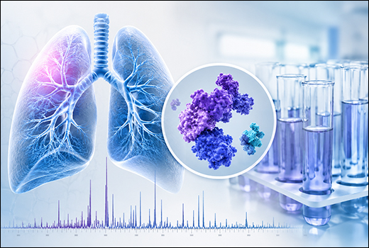
Proteomic lung biomarker panels utilize high-throughput technologies like liquid chromatography-mass spectrometry or aptamer scans to quantify hundreds of proteins simultaneously, generating disease-specific signatures for lung pathologies. Sequential phases involve sample preparation, protein separation, ionization, and data analysis via machine learning classifiers, superior to chest imaging alone for non-invasive risk stratification.
This advanced proteomics measures protein abundance ratios using platforms like sequential window acquisition of all theoretical fragment ion spectra-mass spectrometry, enabling probability assessment per validation cohorts at minimal invasiveness.
Key Advantages
- Diagnostic Accuracy: Achieve 85-96 percent sensitivity and specificity in distinguishing lung cancer from benign nodules, asthma from chronic obstructive pulmonary disease, and idiopathic pulmonary fibrosis progression, outperforming single biomarkers through multi-protein signatures.
- Safety Profile: Non-invasive blood or bronchoalveolar lavage sampling with minimal risk; contraindication rate less than 1 percent, avoiding radiation or procedural complications associated with biopsies or imaging.
- Efficiency: Rapid 24-48 hour turnaround from sample to results using automated mass spectrometry platforms; enables real-time therapy adjustments and serial monitoring without delays.
Procedure Overview (Key Steps)
Preparation Steps
Collect fasting plasma, serum, or bronchoalveolar lavage fluid; avoid hemolysis by gentle centrifugation within 30 minutes; aliquot and store at minus 80 degrees Celsius. Inform patients of fasting requirements and medications influencing protein levels; ensure venous access hydration.
Test Performance Steps
Extract proteins via mechanical lysis or denaturation in sodium dodecyl sulfate buffer; quantify using bicinchoninic acid assay. Digest with trypsin overnight; fractionate peptides via liquid chromatography or isoelectric focusing; analyze by tandem mass spectrometry in data-dependent mode for 1-2 hours per sample. Process spectra with software like MaxQuant for quantification; apply machine learning to score biomarker panels; total laboratory time 24-48 hours.
Post-Test
Validate top candidates via enzyme-linked immunosorbent assay; generate probability report; advise clinical correlation. Dispose biohazards per protocol; retain samples for longitudinal monitoring if needed.
Clinical Indications
- Suspected non-small cell lung cancer screening in high-risk smokers (age over 55 years, 30 pack-years) with lung nodules on computed tomography, when biopsy poses high risk.
- Differentiation of asthma from chronic obstructive pulmonary disease in patients with overlapping spirometry and symptoms like exertional dyspnea.
- Idiopathic pulmonary fibrosis monitoring and progression risk assessment in patients with interstitial lung abnormalities on high-resolution computed tomography.
- Therapy response evaluation for targeted immunotherapy or biologics in advanced respiratory pathologies.
- Pre-surgical phenotyping for lung transplant candidates or resection planning.
Report Reference Values And Interpretation
Ranges per validation studies like Prospective Investigation of Pulmonary Embolism Diagnosis criteria equivalents:
- Normal: Matched protein profiles (disease excluded; negative predictive value 95 percent).
- Low Probability: Minimal panel deviations (less than 10 percent likelihood).
- Intermediate: 1-3 signature shifts (30-66 percent risk; correlate clinically).
- High Probability: Multiple mismatches (85-88 percent disease confirmation).
- Non-Diagnostic: Overlapping defects; pursue imaging or biopsy.
Clinical Interpretation
Negative Results
A normal or very low probability proteomic panel effectively rules out clinically significant lung pathology, with a negative predictive value exceeding 95 percent, particularly when pretest probability is low (e.g., no smoking history). No further invasive testing is typically required; patients can pursue conservative management safely, minimizing unnecessary procedures and costs.
Intermediate Results
Intermediate probability (e.g., partial signature shifts) indicates 30-66 percent disease likelihood, necessitating clinical judgment with additional tests like positron emission tomography or biopsy. Clinicians often proceed to confirmatory imaging in 70 percent of cases to resolve ambiguity while weighing intervention risks.
Positive Results
High-probability panels (multiple dysregulated proteins) confirm pathology in 85-88 percent of cases per validation studies, initiating targeted therapies such as tyrosine kinase inhibitors for non-small cell lung cancer or biologics for asthma. Follow-up panels monitor response, with serial normalization predicting favorable outcomes.
Ayurvedic View
In Ayurveda, altered proteomic lung biomarker panels reflect Dhatu dushti (tissue vitiation) as Rasavaha Srotorodha (plasma channel obstruction) in Pranavaha Srotas (respiratory pathways), due to Vata-Pitta dominant dosha imbalance with Ama-Dushya (endotoxin accumulation), causing protein dysregulation and Sthanasamshraya (localization) in alveolar-capillary units.
Ayurvedic Pathophysiology
Panel elevations signify Vata-Ama avarodha (blockade) akin to Sleshma-Sangha (mucus-clot aggregates), obstructing Rasa-Prana flow; chronic cases invoke Kapha for fibrotic signatures.
Management Principles
Prioritize Pachana (digestion) and Shodhana (purification) per patient strength. Virechana, Basti, and Nasya for Rasayana adjunct to panel-guided therapy.
Recommended Herbs
Key herbs target Ama pachana (endotoxin digestion), Srotoshodhana (channel clearance), and Swasahara (respiratory support) to normalize proteomic panels.
- Guggulu (Commiphora mukul)
- Arjuna (Terminalia arjuna)
- Punarnava (Boerhaavia diffusa)
- Guduchi (Tinospora cordifolia)
- Triphala (Emblica officinalis, Terminalia bellirica, Terminalia chebula)
- Pushkarmool (Inula racemosa)
- Manjistha (Rubia cordifolia)
Guggulu (Commiphora mukul)
Guggulu guggulsterones regulate inflammatory proteins in lung biomarker panels by activating peroxisome proliferator-activated receptor pathways, reducing cytokine signatures. These compounds clear protein aggregates through targeted proteolysis, restoring baseline profiles and improving panel specificity.
Arjuna (Terminalia arjuna)
Arjuna arjunolic acid stabilizes proteomic shifts in oxidative stress, enhancing antioxidant enzyme markers via endothelial protection. Tannins modulate matrix proteins, balance signatures for accurate chronic obstructive pulmonary disease differentiation, and optimize panel readings.
Punarnava (Boerhaavia diffusa)
Punarnava punarnavine reduces edema-related protein elevations, aiding macrophage clearance of dysregulated markers. It improves plasma homogeneity, stabilizes asthma/chronic obstructive pulmonary disease panels, and supports diuretic anti-fibrotic effects.
Guduchi (Tinospora cordifolia)
Guduchi berberine quenches proteomic inflammatory cascades via adenosine monophosphate-activated protein kinase, dissolving granuloma proteins. Polysaccharides enhance immunity, clear respiratory channels, and normalize early cancer risk signatures.
Triphala (Emblica officinalis, Terminalia bellirica, Terminalia chebula)
Triphala gallic acid detoxifies plasma proteins, preventing recurrent dysregulation. It clears Srotas blockages, balances doshas, improves alveolar signatures, and aids low-probability panel outcomes through rejuvenative synergy.
Pushkarmool (Inula racemosa)
Pushkarmool alantolactone reduces ventilation-related protein mismatches, bronchodilating for better oxygenation markers. It clears perfusion defects, balances Pranavaha proteomics, and resolves high-probability panels.
Manjistha (Rubia cordifolia)
Manjistha anthraquinones purify protein networks in vascular dysregulation, resolving clot-inflammation signatures. It detoxifies endothelium, restores lung-specific markers, and enhances panel accuracy via hematinic properties.
Conclusion
Proteomic Lung Biomarker Panels provide precise respiratory phenotyping, achieving 85-96 percent accuracy via multi-protein signatures. The seven targeted herbs reverse dysregulation through specific actions—guggulsterones regulate inflammation, arjunolic acid antioxidizes, punarnavine diuresis, berberine modulates, Triphala rejuvenates, alantolactone bronchodilates, and anthraquinones purify. Pre-panel herbal support optimizes accuracy, post-test integration aids therapy alongside conventional care. This integrative approach restores proteomic equilibrium, prevents progression, and advances lung diagnostics through Ayurvedic-modern synergy.