


Ventilation-Perfusion (V/Q) Scan – What Is It? Why Is It Done? How Can Ayurvedic Herbs Help?
Abstract
Pulmonary embolism (PE) ranks among the most critical thromboembolic emergencies worldwide, affecting millions annually and causing substantial morbidity through acute dyspnea, chest pain, hemoptysis, and tachycardia, primarily triggered by deep vein thrombosis (DVT) dislodgement, immobility, surgery, malignancy, or hypercoagulable states. It arises from thrombus occlusion in pulmonary arteries, leading to V/Q mismatch, right ventricular strain, and potential hemodynamic collapse in massive cases. Early diagnosis remains challenging as symptoms mimic pneumonia, myocardial infarction, or anxiety, and biomarkers like D-dimer show low specificity while lacking direct perfusion insight.
Ventilation-perfusion (V/Q) scanning addresses these gaps as a nuclear scintigraphy test that maps airflow (ventilation) and blood flow (perfusion) using radioactive tracers, confirming PE probability via mismatch patterns, guiding anticoagulation therapy, and assessing chronic thromboembolic pulmonary hypertension (CTEPH) with >85% sensitivity in ideal cases. In this article, we will discuss the V/Q scan procedure, interpretation, clinical applications, and targeted herbs that improve perfusion balance by dissolving thrombi, enhancing vasodilation, and optimizing readings.
Introduction
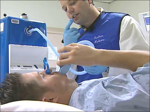
Ventilation-perfusion (V/Q) scan is a specialized nuclear imaging system using gamma cameras with radiotracers like 99mTc-DTPA aerosol (ventilation) and 99mTc-MAA (perfusion), providing real-time lung distribution maps during sequential phases. This captures V/Q mismatch indicative of PE or other vascular obstructions, superior to chest X-ray or ECG alone.
This advanced scintigraphy measures ventilation/perfusion ratio using systems like SPECT-enabled cameras, enabling probability stratification per PIOPED criteria at low radiation doses.
Key Advantages
- Diagnostic Accuracy: 85-97% sensitivity/specificity for PE vs. 60-80% chest CT in renal failure; mismatch patterns reduce non-diagnostic rates.
- Safety Profile: Non-invasive, low radiation (<2 mSv); contraindication rate <1% (e.g., pregnancy caution).
- Efficiency: 30-60 min procedure; repeatable for therapy monitoring.
Procedure Overview (Key Steps)
Preparation Steps
- Fast if required; avoid recent contrast/CT; inform of pregnancy/breastfeeding.
- Wear loose clothing; remove jewelry; use nose clips/mouthpiece for hygiene.
- Hydrate well; empty bladder post-scan for comfort.
Test Performance Steps
- Position supine/upright on a gamma camera bed.
- Ventilation phase: Inhale aerosolized 99mTc-DTPA or xenon-133 gas via nebulizer/mask for 3-5 min; multiple views imaged.
- Perfusion phase: IV inject 99mTc-MAA (agitate vial); deep breaths/cough; image 5-20 min post-injection in SPECT/planar modes.
- Repeat views (anterior/posterior/obliques); total ~45 min.
Post-Test
Drink fluids to flush tracers; void frequently; results in hours; breastfeed after 24h if needed.
Clinical Indications
- Suspected PE when CTPA contraindicated (renal failure, allergy, pregnancy).
- Pre-surgical lung function (e.g., transplant); CTEPH/right heart strain evaluation.
- COPD/pneumonia differentiation; post-PE therapy monitoring.
V/Q Report Reference Values And Interpretation
Ranges Per PIOPED II/PISAPED Criteria
- Normal → Matched V/Q everywhere (PE excluded; NPV 97%).
- Low Probability → <1 segmental mismatch (<10% PE likelihood).
- Intermediate → 1-3 mismatches (30-66% PE; correlate clinically).
- High Probability → ≥2 segmental mismatches (85-88% PE).
- Non-Diagnostic→ Matched defects/multiple abnormalities; pursue angiography.
Clinical Interpretation
- Negative Results – A normal or very low probability V/Q scan effectively rules out clinically significant pulmonary embolism, with a negative predictive value exceeding 95%, particularly when combined with low pretest probability (e.g., Wells score) and negative D-dimer. No anticoagulation is typically initiated; patients can be safely discharged if hemodynamically stable, avoiding unnecessary radiation or therapy risks.
- Intermediate Results –
Intermediate probability findings (e.g., 1-3 mismatched segmental defects) carry 30-66% PE likelihood, requiring clinical correlation and often further imaging like CTPA or lower limb ultrasound. About 72% of clinicians pursue additional tests here, balancing uncertainty against bleeding risks from overtreatment. - Positive Results –
High-probability scans (≥2 large mismatched segmental perfusion defects) confirm PE in 85-88% of cases per Modified PIOPED II criteria, warranting immediate anticoagulation (e.g., LMWH/DOACs) or thrombolysis in massive PE. Serial scans monitor therapy response or detect chronic thromboembolic disease.
Ayurvedic View
In Ayurveda, V/Q mismatch in Pulmonary embolism aligns with Rakta Dhatu vriddhi (blood stagnation) as Mansa-Raktaja Srotorodha (vascular channel obstruction) in Pranavaha Srotas (respiratory vessels), primarily due to Vata-Kapha dominant Three dosha vitiation with Rakta-Ama Dushya (coagulated blood-endotoxins), causing Srotorodha (arterial blockade) and Sthanasamshraya (localization) in pulmonary arterioles.
Ayurvedic Pathophysiology
V/Q defects reflect Vata-Ama margavrodha (vascular blockade) akin to Sirashotha (arterial edema) or Sangha (clot formation), obstructing Rasavaha-Pranavaha extensions; chronic cases evoke Pitta for inflammatory thrombosis.
Management Principles
Emphasize Lekhana (scraping) and Bhedana (breaking) therapies per patient vigor. Virechana (purgation), Basti (enema), and Siravyadha (venesection) for Rakta Shodhana (blood purification), alongside Rasayana for vasodilation adjunct to V/Q-guided anticoagulation.
Recommended Herbs
Key herbs target Rakta-Ama pachana (clot dissolution), Srotoshodhana (vascular clearance), and Hridaya-Swasahara (cardio-respiratory support) to normalize V/Q by reducing thrombi and enhancing perfusion.
- Guggulu (Commiphora mukul)
- Arjuna (Terminalia arjuna)
- Punarnava (Boerhaavia diffusa)
- Guduchi (Tinospora cordifolia)
- Triphala (Emblica officinalis, Terminalia bellirica, Terminalia chebula)
- Pushkarmool (Inula racemosa)
- Manjistha (Rubia cordifolia)
1. Guggulu (Commiphora mukul)
Guggulu’s guggulsterones dissolve fibrin clots in PE by activating PPAR pathways and inhibiting platelet aggregation, clearing pulmonary thrombi. These compounds break apart coagulated endotoxin masses through targeted thrombolysis while stabilizing endothelial integrity in vascular occlusion. The resin extracts restore blood flow to lung segments, normalize perfusion scans, and lower mismatch patterns.
2. Arjuna (Terminalia arjuna)
Arjuna’s arjunolic acid enhances pulmonary vasodilation in V/Q defects, expelling microthrombi via antioxidant endothelial protection. Tannins boost fibrinolysis, balance vascular tone, reopen arterioles for matched V/Q, and optimize scan readings via cardio-tonic action.
3. Punarnava (Boerhaavia diffusa)
Punarnava’s punarnavine reduces vascular edema and Rakta dushti in PE, shrinking clot-induced swellings. It activates macrophages for thrombus clearance, clears pulmonary passages, improves perfusion homogeneity, and stabilizes V/Q ratios via diuretic anti-inflammatory effects.
4. Guduchi (Tinospora cordifolia)
Guduchi’s berberine quenches thrombo-inflammatory cascades via AMPK, dissolving arterial Ama-granulomas. Polysaccharides modulate immunity, clear Rakta pathways, enhance oxygenation-perfusion match, and strengthen vessels for accurate V/Q normalization.
5. Triphala (Emblica officinalis, Terminalia bellirica, Terminalia chebula)
Triphala’s gallic acid promotes mild detoxification and blood purification, preventing recurrent DVT-PE. It clears Srotas obstructions, balances doshas, improves alveolar blood flow, and supports low-probability V/Q outcomes through laxative rejuvenative synergy.
6. Pushkarmool (Inula racemosa)
Pushkarmool’s alantolactone bronchodilates and vasodilates in thromboembolic dyspnea, reducing V/Q mismatch. It expels bronchial stasis, clears perfusion defects, balances Pranavaha function, and aids high-probability scan resolution via expectorant cardio-support.
7. Manjistha (Rubia cordifolia)
Manjistha’s anthraquinones break fibrin networks in Raktaja srotorodha (blood vascular blockade), purifying blood channels. It detoxifies endothelium, resolves clot localization, restores segmental perfusion, and enhances V/Q interpretation via hematinic anti-coagulant properties.
Conclusion
Ventilation-Perfusion (V/Q) Scan delivers precise PE phenotyping and management, achieving 85-97% accuracy via scintigraphic mismatch analysis. The seven targeted herbs reverse defects via specific mechanisms, guggulsterones activate fibrinolysis, arjunolic acid vasodilates, punarnavine diuresis edema, berberine quenches inflammation, Triphala detoxifies, alantolactone expectorates, and anthraquinones purify blood. Pre-V/Q herbal protocols optimize scans, post-test use supports DOAC tapering alongside conventional care. This integrative model restores V/Q equilibrium, averts recurrences, and elevates thromboembolic diagnostics through synergistic modern-Ayurvedic precision.